


1. Introduction
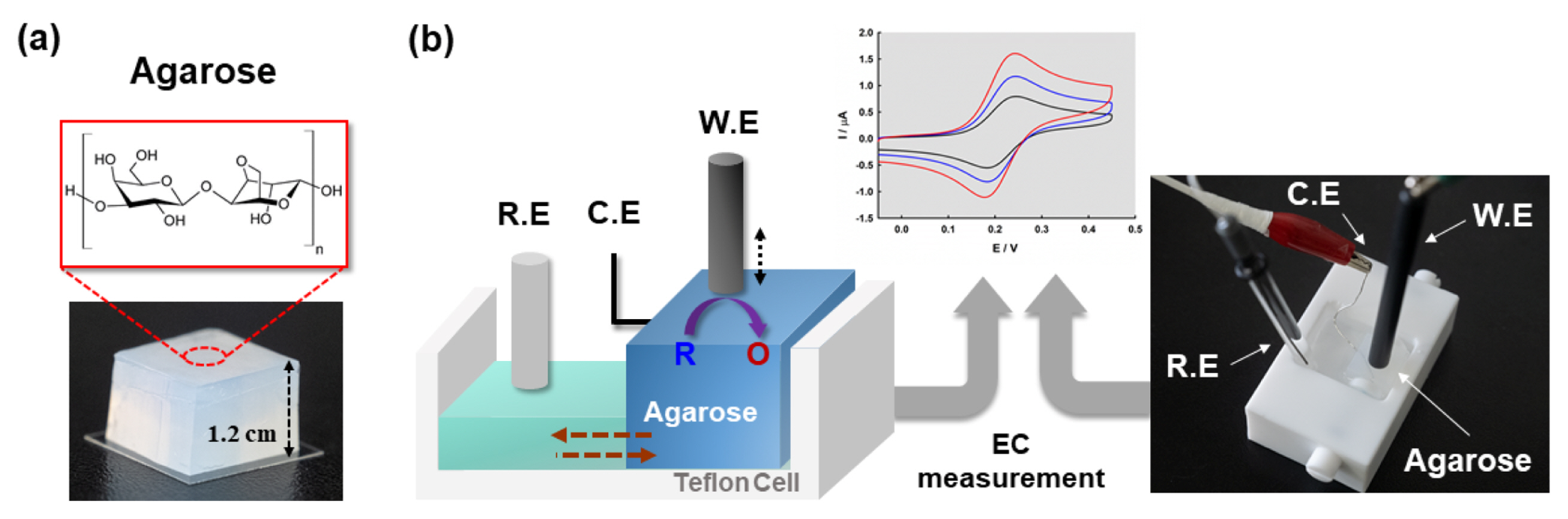
Hydrogels are three-dimensional (3D) materials in which a network of hydrophilic polymer chains holds a large quantity of water [1]. The specific structures and biocompatible properties [2] allow application in many research fields, such as biosensors [3–5], tissue engineering [6], biomedical engineering [7], and drug delivery [8, 9]. Also, hydrogels as solid electrolytes are an emerging material for energy storage and conversion fields due to their high structural flexibility [10], electronic conductivity [11], and electrolyte permeability [12]. Among the types of hydrogels, agarose is a widely used material due to its inexpensive, non-toxic, easy synthesis and good experimental properties. Agarose is a linear polysaccharide consisting of 1,3-linked β-D-galactopyranose and 1,4-linked 3,4-anhydro-α-L-galactopyranose as a repeating unit. Agarose gels have a wide distribution of pore-size ranging from 1 to 900 nm depending on agarose composition [13] and can be characterized by transmission electron microscopy (TEM) [14] and atomic force microscopy [15].
For the numerous applications in electrochemistry, understanding the diffusion properties of redox molecules in the gel and the interaction with the gel network are important, but few studies have investigated this aspect compared to the study of electrochemistry in the bulk solution phase [16–18]. In previous research, mass transport properties of various solutes in agarose gel have been studied in terms of steric, electrostatic, and chemical interactions using fluorescence correlation spectroscopy (FCS) [19]. In another study, Yin et al. investigated the intermolecular interaction between electroactive probes and the microdomain in polysaccharide hydrogels using a voltammetric technique and showed variations in the net shift of normal potentials and the ratio of binding constants (Kb) of redox molecules in hydrogels [20]. Recently, Hwang et al. introduced a hydrogel pen (HYPER) for electrochemical reaction between pyramidal-shaped agarose and a macroelectrode by controlled contact area. They reported that the mass transport properties in agarose gel were similar to those in solution except for a slight decrease in diffusion coefficient [21].
The basic techniques of electrochemistry such as cyclic voltammetry (CV) [22,23] or chronoamperometry (CA) [24] were interpreted by the Randles-Sevcik equation, Cottrell equation, or Shoup and Szabo equation [25], which are based on diffusional mass transport without migration or convection [26–30]. Migration can be ignored with excess electrolyte, but poor reproducibility and deviation from the fundamental theory of electrochemistry can occur by uncontrollable natural convection. Aoki et al. have demonstrated a method to suppress natural convection by enhanced viscosity of the solution using sodium alginate (SA) without any change of diffusion properties of the redox molecule [31]. Recently, the effects of an active vibration isolator (AVI) and the electrode position in the cell were studied to reduce natural convection for slow scan voltammetry [32].
In this report, the purpose of our work is to study the mass transport properties of FcMeOH as a solute at the agarose hydrogel interface depending on the gel concentration. Previous reports have investigated the diffusion phenomena in the gel or gel/solution interface [33], but only rare studies exist for electrochemical measurement of the diffusional properties of the solute at the gel/electrode interface. Also, a gel interface with locally denser structure than the bulk gel can reduce the natural convection due to enhanced interaction between the agarose polymer network and solute. An Au disk electrode and Fe(CN)64− as the redox molecule were used to confirm the reduced natural convection on the gel interface using slow scan voltammetry. The simple setup with a home-made cell and electrode configuration is shown in Scheme 1.
2. Experimental
2.1 Materials
Agarose (Low EEO), ferrocenemethanol (FcMeOH) (97%), potassium chloride (99.9%), and potassium hexacyanoferrate(II) trihydrate (99.9%) were obtained from Sigma-Aldrich (St. Louis, MO). A gold disk electrode (0.8 mm radius) was purchased from BAS Inc. MicroPolish alumina (0.3 μm, 0.05 μm) powders and a polishing micro-cloth pad were prepared and obtained from Buehler. All the chemicals and reagents used in this work were of reagent grade, and water (>18 Mω·cm) was obtained from a Millipore Milli-Q purification system.
2.2 Preparation of agarose hydrogel for electrochemistry
Agarose solutions with various concentrations (11 w%, 6.6 w%, 4.8 w% agarose in water) were prepared in a microwavable cylinder-shaped container equipped with a sealing cap. The solution in the container was preconditioned in a 90°C water bath for 1 hour until the agarose was completely dissolved. Air bubbles, which will disrupt the gel, were eliminated by heating under microwave (700 W) power for 30 s until the solution became viscous and transparent. The prepared hot agarose solution was poured into a glass mold and slowly cooled in a humidity chamber. The solidified agarose was cut into desired sizes, carefully separated from the glass mold, and stored in distilled water. Before electrochemical measurement, the agarose gels were immersed in aqueous solution of redox molecules and supporting electrolyte for 8 h for complete equilibrium. For electrochemical measurement, redox material containing agarose gel was installed in a home-made cell.
2.3 Electrochemical measurement
The surface of the working electrode was polished with alumina paste on a wet cloth and was rinsed with distilled water in an ultrasonic bath. Ag/AgCl and Pt wire (0.5 mm diameter) were used as a reference electrode and a counter electrode, respectively. Electrochemical experiments were carried out using a CHI 601e potentiostat (CH Instruments, Austin, TX), and an Au disk electrode was fixed vertically with a clamp and support jack. Prior to the electrochemical measurement, the hydrogel pad was placed in the home-made cell, and the Pt wire was positioned inside the agarose gel. A digital microscope (AM3113, Dino-Lite) was used to confirm contact between the agarose gel and working electrode.
3. Results and Discussion
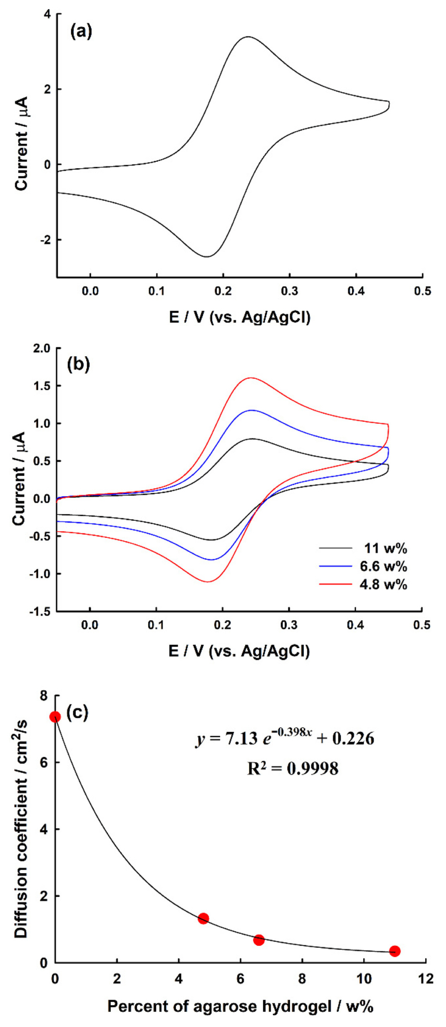
In order to analyze how the diffusion changes for redox species in the agarose hydrogel, ferrocenemethanol (FcMeOH) was selected as an electrochemical redox material. FcMeOH is a representative redox molecule that performs a one-electron transfer and shows a good reversible electrochemical reaction, which is suitable for observing redox diffusion in an agarose hydrogel. Here, 0.1 M KCl was used as an electrolyte to screen the migration effect. The CV of FcMeOH measured on the Au disk electrode in the solution phase is shown in Fig. 1(a). The measured anodic peak potential (Epa) and cathodic peak potential (Epc) were ca. 237 and ca. 176 mV, respectively, and the peak-to-peak separation (ΔE) was ca. 61 mV. This value is similar to the theoretical ΔE value of a one-electron reversible system (59 mV at 25°C). Here, the diffusion coefficient of redox (i.e., FcMeOH) can be obtained using the Randle-Sevcik equation at room temperature [34].
Where Ip is the net peak current (A), n is the number of electron transferred in the redox event, A is the area of the electrode (cm2), D is the diffusion coefficient of redox (cm2/s), C is the bulk concentration of redox (mol/cm3), and v is the scan rate (V/s). The diffusion coefficient of FcMeOH obtained from Eq. (1) is 7.36 × 10⁻⁶ cm2/s. Based on this value, diffusion coefficients of FcMeOH were compared under various agarose concentrations.
To assess the diffusional properties of the redox molecule on the agarose surface, the CV of FcMeOH was measured according to concentration of agarose (Fig. 1(b)). As the concentration of agarose decreased to 11.0, 6.6, and 4.8 w%, the Ip values were measured to be 0.710, 0.995, and 1.39 μA, respectively. The diffusion coefficients (DFcMeOH) obtained from Eq. (1) are 0.344 × 10⁻⁶, 0.677 × 10⁻⁶, and 1.32 × 10⁻⁶ cm2/s, respectively. The DFcMeOH values on the agarose surface were nearly the same order of magnitude as those in the solution, but mass transport properties of solutes were reduced on the gel interface. The structure of the agarose surface is different from that in the bulk gel due to a dense fiber concentration or entangled fiber organization [29]. Therefore, the permeability of the solute in the interface decreases as the concentration of agarose increases, assumed to be caused by steric hindrance between solute and agarose polymer fibers. As the concentration of the agarose hydrogel increased, the diffusion coefficient of redox decayed exponentially, which agrees with the mass transport properties of small molecules in the hydrogel (Fig. 1(c)) [35, 36].
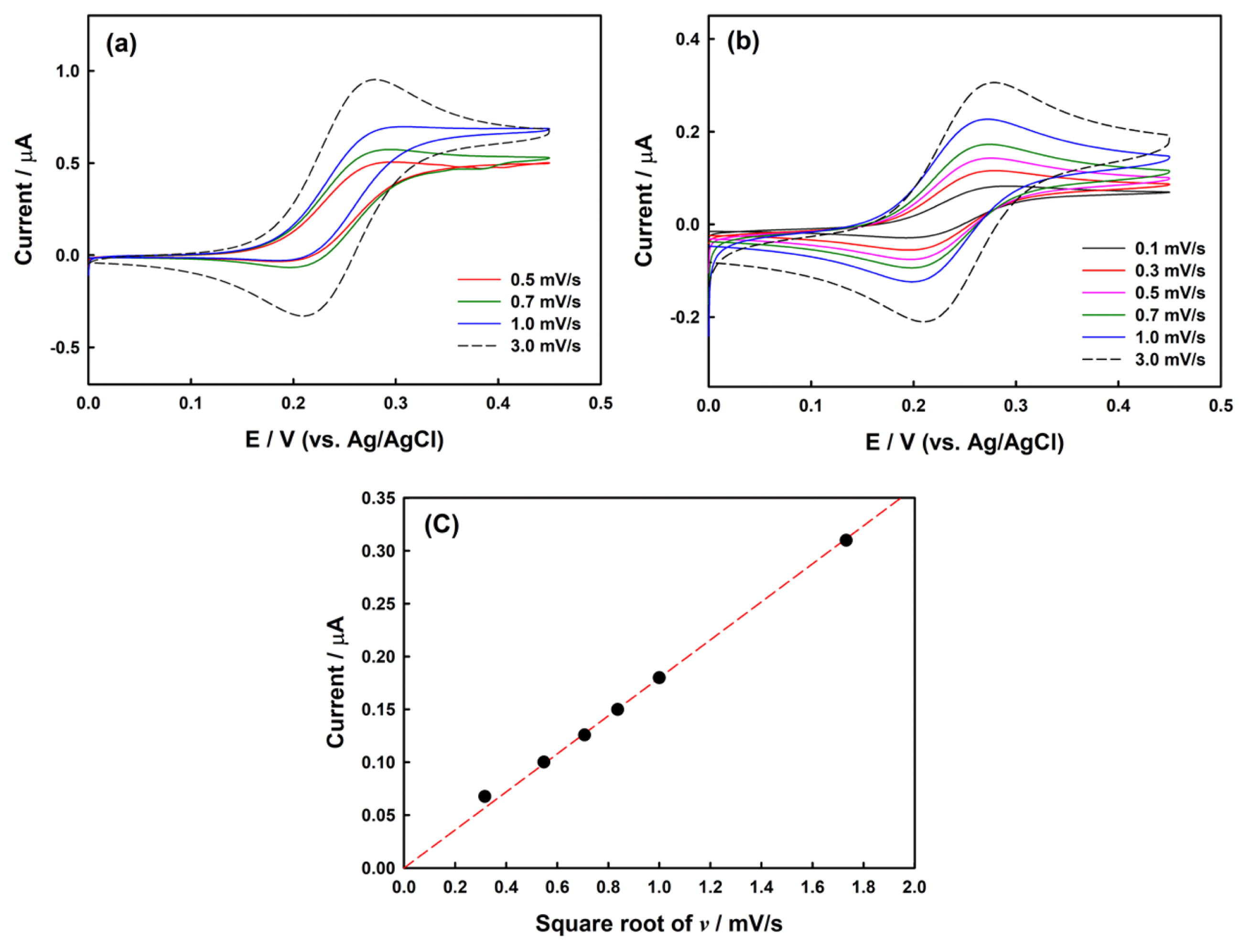
We also investigated whether there was change in the diffusion coefficient of redox when the scan rate was changed in the agarose hydrogel condition. To confirm this, CVs were measured while increasing the scan rate to 0.01, 0.02, 0.05, 0.1, and 0.2 V/s under 11, 6.6, and 4.8 w% agarose hydrogel conditions. As shown in Fig. 2, the peak current also increased as the scan rate increased, and a linear relationship was established by plotting the square root of the scan rate and peak current. Linearity of the square root of the scan rate and the peak current is observed in electrochemical reactions where mass transport is mainly governed by diffusion [30]. Therefore, regardless of the concentration of agarose hydrogels, the main factor of mass transfer is diffusion, and other factors (i.e., convection and migration) are negligible under the normal scan rate condition.
Mass transport, especially the diffusion process, is a key step in electrochemical analysis since it allows analytes to transport from bulk solution to the electrode. The agarose polymer backbone is composed of various ionic chemical moieties such as sulfonate, ester sulfate, ketal pyruvate, and carboxylic groups, which can affect the diffusion or convection properties of solutes in gel by steric, specific, and electrostatic interactions [19]. These interactions between the solute and gel network will cause not only a previously resultant diffusive hindrance, but also convective hindrance.
To investigate the influence of a hydrogel to reduce natural convection, CVs were obtained in the solution phase and on the agarose surface, respectively. As shown in Fig. 3 (a), conventional CVs were obtained at a scan rate lower than 3.0 mV/s. Decreasing the scan rate from 1.0 to 0.5 mV/s resulted in sigmoidal shapes rather than the peak shapes assumed to be due to natural convection and also showed less reproducibility of current at the steady-state region [33]. Unpredictable current by natural convection was observed with a scan rate as low as 0.1 mV/s on the agarose surface (Fig. 3(b)) for up to 2.5 hours with long-term electrolysis. The plot of peak current against v1/2 (Fig. 3(c)), as predicted by Eq (1), showed a fine linear relationship down to 0.3 mV/s without natural convection. From these voltammetric results, natural convection can be reduced using agarose hydrogel as the solid electrolyte, probably due to various interactions between the solute and ionic agarose polymer network.
4. Conclusions
The mass transport properties of solute on agarose hydrogel interface were investigated using CV. The CVs with various compositions of agarose showed hindered diffusional properties of FcMeOH at the gel interface compared to the bulk solution, but diffusion-controlled current involved negligible other mass transport factors (i.e., convection and migration). Also, slow scan voltammetry with agarose hydrogel revealed minimized natural convection down to scan rates of 0.3 mV/s without any complicated equipment. These results suggest that the hydrogel as a solid electrolyte can be a good candidate for slow scan voltammetry or in long-time chronoamperometric measurement.