


1. Introduction
Listeria monocytogenes (LM) is a foodborne pathogen that widely exits in water, soil, plants, feces, and it can arouse several diseases, including abortion, meningitis and septicemia, to the humans with high morbidity and fatality rate by contaminating various food [1ŌĆō4]. Owing to its great endanger, LM was listed as one of the most hazardous and deadliest pathogens [5,6]. Consequently, there have been several interesting electrochemical sensors, such as DNA-based sensor [3], sandwich immunosensor sensor [5,7] and peptide-based sensor [4], developed for the detection of LM since the electrochemical method is simpler, more sensitive, easy to operate, time-saving and low-cost compared to the other methods [8ŌĆō10]. ThereŌĆÖs no doubt that these sensors have considerably great applications, while the requirement of DNA, antibody, or aptamer endows the sensing processes with complex operation and high cost [11].
Molecularly imprinted polymer (MIP) is a tailor-synthesized receptor that can display specific and complementary recognition sites towards the related templates [12ŌĆō15]. Compared to the other receptors including antibodies, DNA and aptamers, MIP has several unique superiorities, such as cost-effective, high stability and simple preparation [16ŌĆō18]. Over the past decades, MIP has been confirmed to show great application values in the detection of protein, virus, and many organic molecules [19ŌĆō21]. In recent years, there are several MIP-like sensors developed for pathogen detection [22,23], this inspired us that it may be a perfect proposal to design a MIP-like electrochemical sensor for LM detection.
Selection of optimal carrier in preparing MIP is very important for the improvement of sensitivity. MXene, discovered and studied extensively in 2011, is a newly emerging transition metal carbide and/or nitride material and has been widely in various fields such as energy conversion and storage, field effect transistors and separation catalysis [24ŌĆō28] as well as microbial electrochemical systems [29ŌĆō32]. The general formula for MXene is Mn+1XnTx (e.g., Ti3C2Tx), where M denotes early transition metal and X represents carbon or nitrogen, while Tx refers surface functional groups (e.g., -OH, -O, -F) [33ŌĆō37]. Because of its huge specific active area, good conductivity and mechanical properties, MXene has great application prospects as a promising electrode material for electrochemical sensors [38ŌĆō41]. As for the MIP synthesization, there are several approaches, including electrochemical polymerization, thermal, and optical methods, have been proposed, among which, the first method has attached the most considerable attentions since it offers a very simple and controllable modification of the polymer to the electrode surface [42ŌĆō45]. Therefore, the utilization of suitable electroactive functional monomers is another key step for the preparation of MIP-based sensor. Thionine (Th), an aromatic dye with two amino groups, has demonstrated to be a desirable candidate as a promising functional monomer since its electropolymerization can generate a poly(thionine) film with superior electron transfer capability, and it can also be served as a signal molecule with good stability and electrochemical response [46,47].
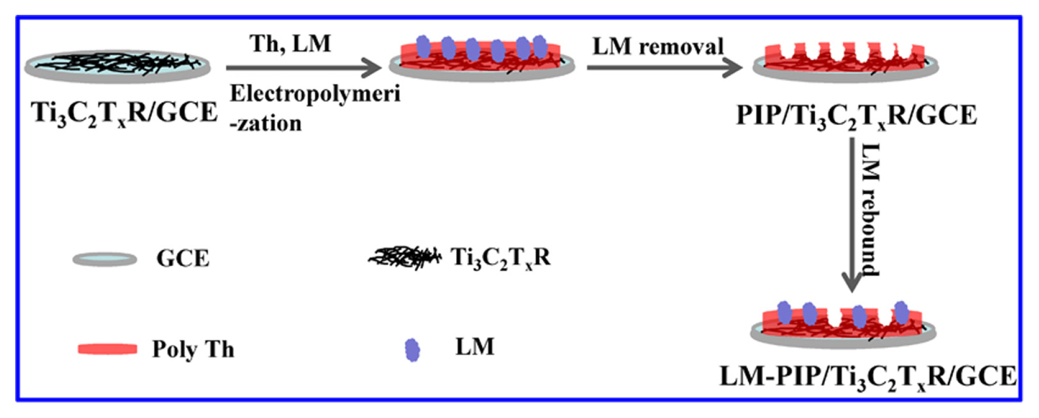
Based on the above statements, in this work, a highly sensitive MIP-like (pathogen imprinted polymer, PIP) integrated probe electrochemical sensor was proposed for LM detection by preparing Ti3C2Tx MXenes nanoribbon (Ti3C2TxR) as the carrier of PIP and using Th acted simultaneously as the functional monomer and signal probe (Scheme 1). After optimizing three key experimental factors, a considerably low analytical limit and wide linearity of LM were obtained. This work offers a simple and low-cost pathway for the electrochemical detection of LM.
2. Experimental
2.1 Preparation of Ti3C2TxR
Ti3C2TxR was prepared according to the previous works with similar modification [48]. In brief, 1.0 g Ti3AlC2 was added into 30.0 mL hydrofluoric acid solution and stirred gently at 40┬░C to form Ti3C2Tx nanosheets (Ti3C2TxS) that can be collected after centrifugation and washed with water. Next, the obtained Ti3C2TxS powder of 0.2 g was mixed with 25.0 mL KOH solution (6.0 M) and further stirred under N2 atmosphere at 25┬░C for four days, thus resulting the information of Ti3C2TxR.
2.2 Construction of PIP sensor
Firstly, 2.0 mg Ti3C2TxR powders were dispersed into 4.0 mL water and treated for 10 min with ultrasound. Next, 6 ╬╝L of the produced Ti3C2TxR dispersion was dripped onto the GCE surface and dried with infrared lamp to form Ti3C2TxR/GCE. Then, the PIP sensor was prepared by electropolymerizing of 5.0 mM Th and LM on the Ti3C2TxR/GCE surface in 0.1 M PBS (pH 6.0) for 20 cycles with the potential range from ŌłÆ0.4 to 0.4 V at a scan rate of 50.0 mV/s. Afterwards, the polymer modified electrode was soaked in 0.5 M hydrochloric acid solution to remove the LM template, which was labeled as PIP/ Ti3C2TxR/GCE. As a comparison, non-pathogen imprinted polymer (NIP) modified Ti3C2TxR/GCE (NIP/Ti3C2TxR/GCE) was constructed with the similar processes just in the inexistence of LM.
2.3 Detection of LM
Firstly, PIP/Ti3C2TxR/GCE was incubated in LM solution with different concentrations for 20 min to recognize and capture LM, denoting the formed electrode as LM-PIP/Ti3C2TxR/GCE. Next, the differential pulse voltammetry (DPV) test was carried out in 0.1 M PBS (pH 6.0). The absolute value of peak current difference (|ΔIp|) was recorded and calculated according to the following equation:
where
I p 0
3. Results and Discussion
3.1 Characterization of Ti3C2TxR
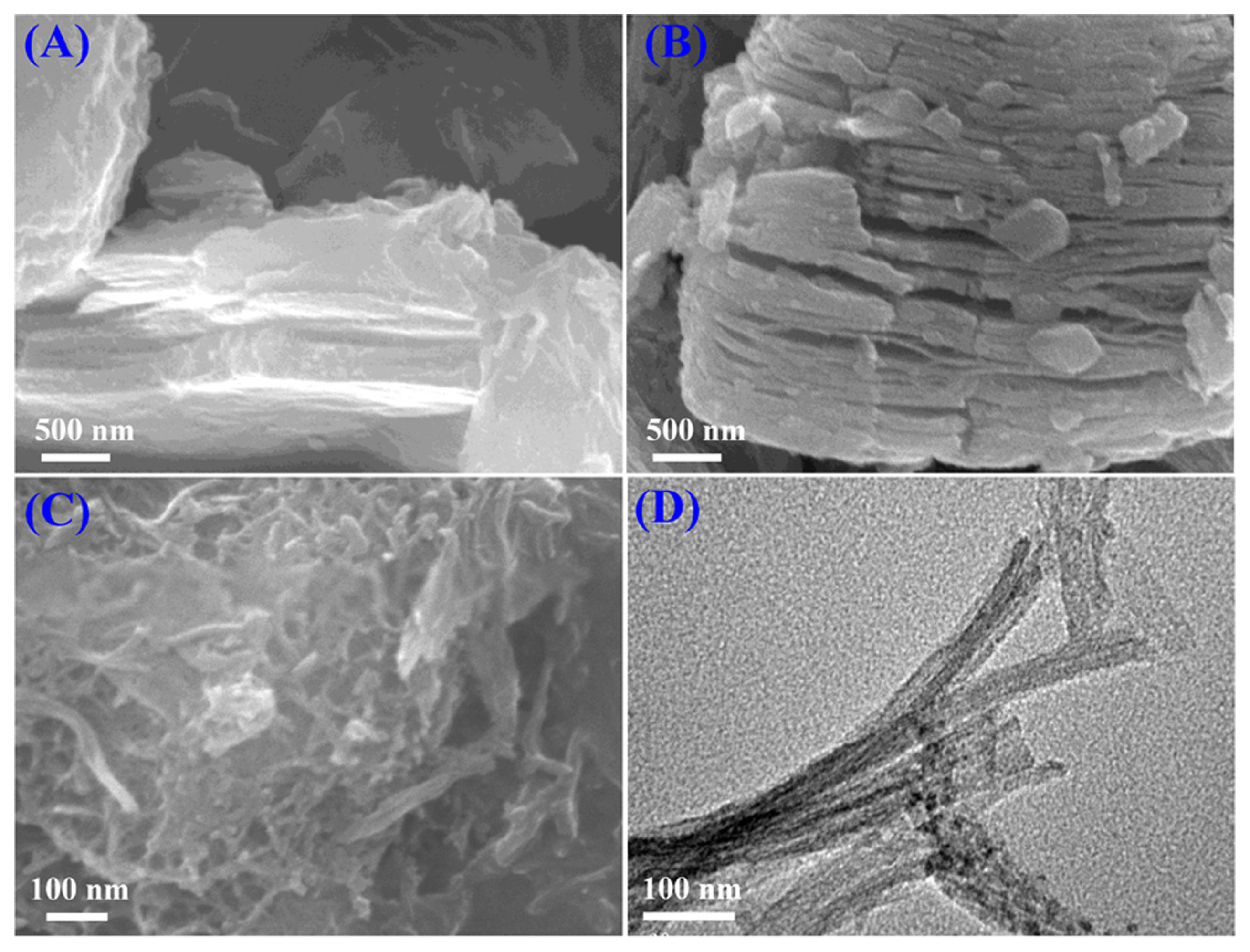
The preparation of Ti3C2TxR was investigated by scanning electron microscopy (SEM) and transmission electron microscopy (TEM). As show in Fig. 1a, the purchased Ti3AlC2 exhibits a closely aligned structure. After etching Ti3AlC2 with hydrofluoric acid, the loosely 2D layered structure can be observed obviously for the formed Ti3C2TxS (Fig. 1b). From the SEM image (Fig. 1c) and TEM image (Fig. 1d) of Ti3C2TxR, it can be found that the nanoribbon structure was obtained for Ti3C2TxR, and the average diameter is about 35 nm, indicating that Ti3C2TxR was produced successfully.
3.2 Electrochemical studies of PIP sensor
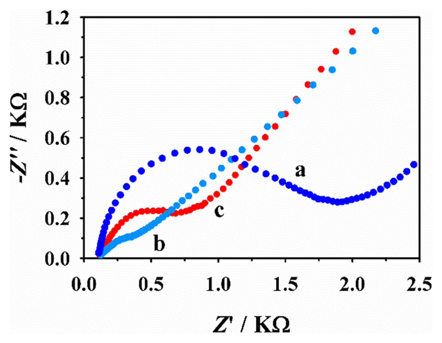
Firstly, the imprinted process of PIP sensor was studied by electrochemical impedance spectroscopy (EIS). As shown in Fig. 2, it displayed the Nyquist diagrams of various electrodes in the solution of 2.0 mM [Fe(CN)6]3ŌłÆ/[Fe(CN)6]4ŌłÆ. Before the removal of LM template, the modified electrode shows a remarkable interfacial resistance, revealing that the presence of LM has larger obstruction to decrease the electron transfer rate and increase the resistance of the electron flow. However, when the LM template was removed from the imprinted cavities, an obvious reduction of interfacial resistance could be found at PIP/Ti3C2TxR/GCE. The reason is that there are a large number of imprinted cavities produced on the electrode surface through the elution of LM template, which increases the diffusion rate of [Fe(CN)6]3ŌłÆ/4ŌłÆ via the PIP film and makes it easier to transfer electron. The impedance changes of the modified procedures also suggested that the PIP sensor has been constructed successfully. Furthermore, after the incubation of PIP/Ti3C2TxR/GCE in the LM solution, an increased impedance can be observed at LM-PIP/ Ti3C2TxR/GCE, this can be attributed to the rebound LM cell in imprinted cavities that can block the channel of [Fe(CN)6]3ŌłÆ/4ŌłÆ to the electrode surface.
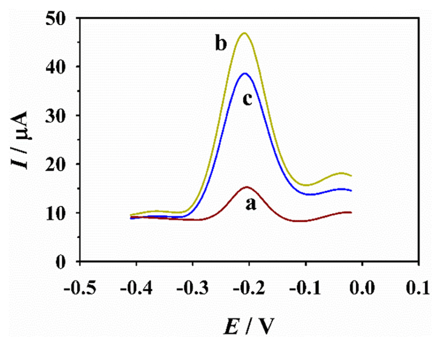
Next, the feasibility of PIP sensor towards LM detection was evaluated by DPV since it can exhibit high sensitivity and resolution. As exhibited in Fig. 3, before the removal of LM template, the as-prepared PIP electrode possesses a weak peak current of Th at ~ŌłÆ0.2 V; along with the LM elution from the electrode surface, the formed imprinted cavities in the polymer layer can generate many electron transmission channels, leading to an increased obviously oxidation signal of Th. Whereas with the incubation of PIP/Ti3C2TxR/GCE in LM solution (103 CFU mLŌłÆ1), the rebound LM cell into the imprinted cavities can impede the electron channel for Th, thus leading to the decrease of Th signal current. As for NIP/ Ti3C2TxR/GCE, there is nearly no difference observed before and after its incubation in LM solution that is because NIP/Ti3C2TxR/GCE cannot recognize and bound LM cells due to the inexistence of LM imprinted cavities. These results proved the designed detection strategy for LM in this work is feasible
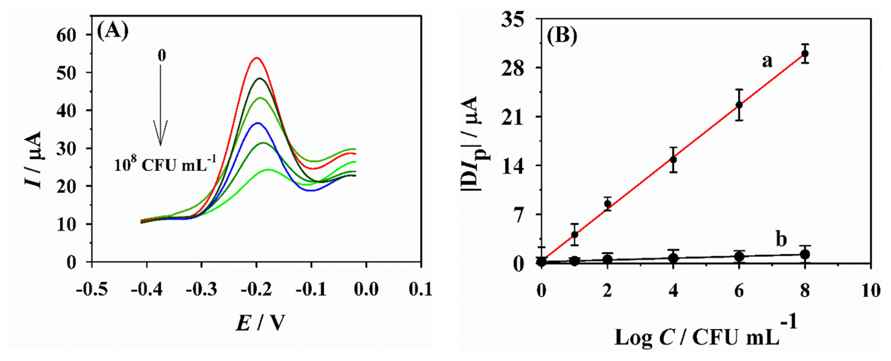
Before the quantitative analysis of LM, several key parameters were optimized for obtaining the best analytical capability of the PIP sensor. As shown in Fig. S1ŌĆōS3, the optimal conditions for the concentration of template LM, the polymerization cycles of Th and the incubation time of PIP/Ti3C2TxR/GCE in LM solution were 2├Ś107 CFU mLŌłÆ1, 18 and 25 min, respectively. Then, under the optimal conditions, DPV was used to monitor the concentration of LM in pH 6.0 PBS. Fig. 4a displayed the DPV responses of LM with various concentrations at PIP/Ti3C2TxR/ GCE. It can be found with the increase of LM concentrations, the peak signals of Th gradually decrease owing to the blocking of the imprinted cavities, and the |╬öIp| values are linear with LM concentrations in a range from 10 to 108 CFU mLŌłÆ1. The corresponding linear regression equation is |╬öIp| (╬╝A) = 0.3714 + 3.706 Log C (R = 0.9984) and the limit of detection (LOD) was obtained to be 2 CFU mLŌłÆ1 (S/N = 3) (Fig. 4b I). For comparison, NIP/Ti3C2TxR/GCE was used also to monitor LM. As shown in Fig. 4b II, in contrast to PIP/Ti3C2TxR/GCE, there is almost no change for the |╬öIp| values with different concentrations of LM, revealing that NIP/Ti3C2TxR/GCE canŌĆÖt bound LM cells due to the lack of specific cavity. Especially, compared to the previous studies (Table 1), the present constructed PIP sensor exhibits a preferable analytical capability with a relatively lower LOD and wider linearity, allowing the highly effective determination of LM.
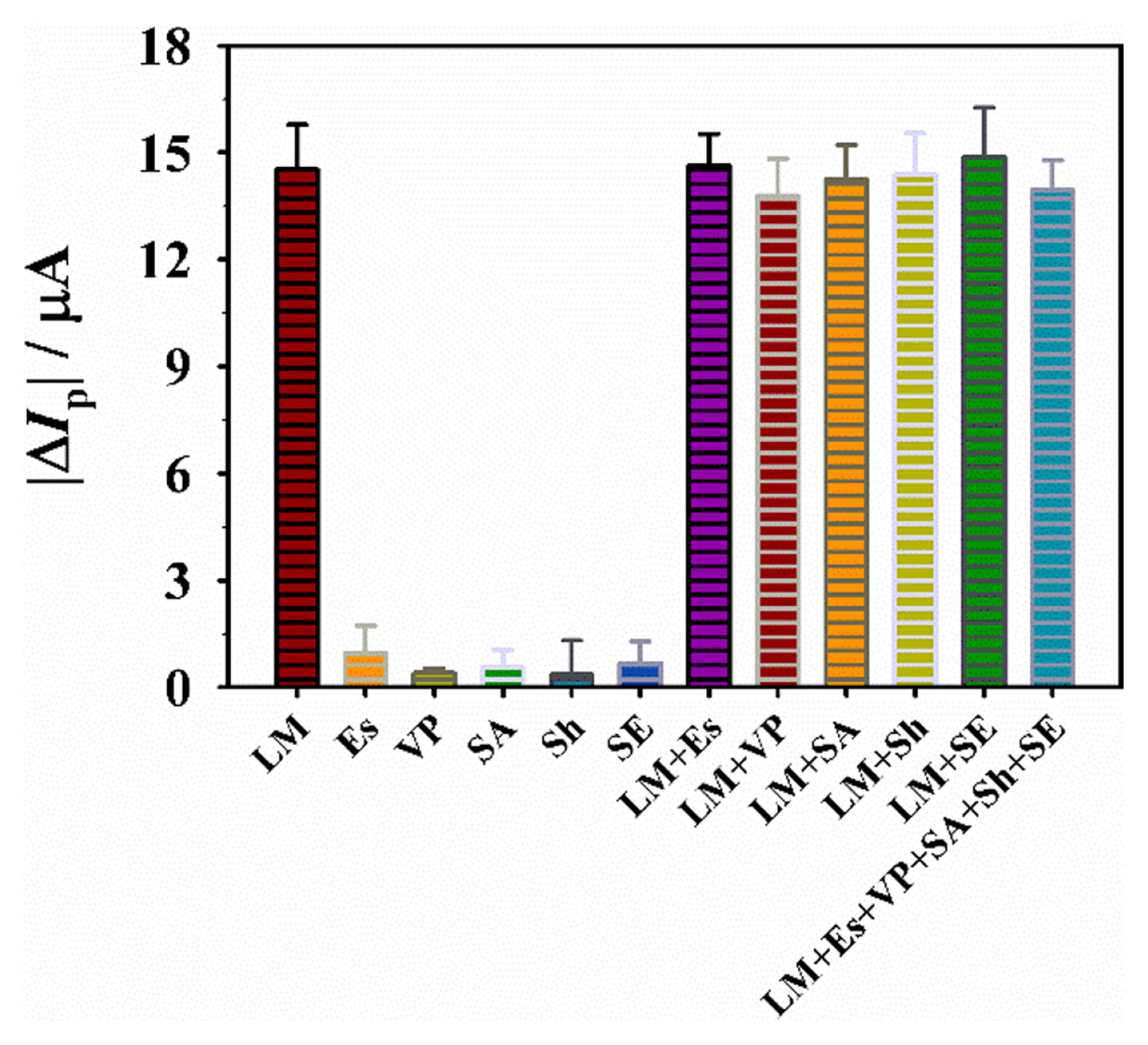
To study the selectivity of the PIP sensor, the asprepared PIP/Ti3C2TxR/GCE was used to detect LM and other interfering pathogens including Escherichia (Es), Vibrio parahaemolyticus (VP), Staphylococcus aureus (SA), Shigella (Sh), and Salmonella enteriditis (SE). Fig. 5 displays the |╬öIp| values of PIP/Ti3C2TxR/GCE towards LM and other pathogens at the same concentration (104 CFU mLŌłÆ1) in 0.1 M PBS (pH 6.0), and the experiments were carried out in triplicate. The results reveal that the presence of LM can cause a significant change (|╬öIp|) in the peak current, while almost no |╬öIp| values were observed for the other pathogens (less than 6.0%). In view of the fact that one sample may contain multiple pathogens in real situation, so the DPV responses of LM in the standard solutions containing other interfering pathogens were studied further. The results reveal the current responses did not change significantly with the |╬öIp| values ranging from 13.78 to 14.62 ╬╝A, indicating that of the established PIP sensor exhibits satisfactory selectivity.
Another two significant indicators for sensor performances are reproducibility and stability. For evaluating the reproducibility of PIP sensor, eight parallel-fabricated PIP/Ti3C2TxR/GCE were applied to record the DPV response of 104 CFU mLŌłÆ1 LM. As showed in Fig. S4a, it can be found that the RSD value of |╬öIp| is only 3.65%, indicating that the asprepared PIP/Ti3C2TxR/GCE offers desirable reproducibility. In addition, the storage stability of the fabricated PIP sensor was investigated through comparing the mean |╬öIp| value using the stored PIP/ Ti3C2TxR/GCE (4┬░C for 28 d) with those of fabricated freshly ones (Fig. S4b). The results show that the PIP sensor can retain 93.2% of its initial signal, revealing a long lifetime for the fabricated sensor.
4. Conclusions
In this study, a pathogen imprinted film fabricated on a Ti3C2TxR modified electrode was developed for the first time for the highly effective determination of LM. In strategical, the introduced Th cannot only act as an effective functional monomer, but also can dedicate as an internal reference probe of the PIP sensing platform. In addition, Ti3C2TxR can offer a desirable carrier with large specific active area and good conductivity. By virtue of the optimal experimental conditions, the fabricated PIP/Ti3C2TxR/GCE sensor displayed a low LOD (10ŌĆō108 CFU mLŌłÆ1), wide linearity (2 CFU mLŌłÆ1), high selectivity, excellent repeatability and stability. This sensitive, simple, and selective sensing strategy for LM opens a new horizon for its sensitive and rapid recognition, which also holds great promise for the future applications to other pathogens.