Simple and Sensitive Electrochemical Sandwich-type Immunosensing of Human Chorionic Gonadotropin based on b-cyclodextrin Functionalized Graphene
Article information
Abstract
The effective detection of human chorionic gonadotropin (HCG) is considerably important for the clinical diagnosis of both of early pregnancy and nonpregnancy-related diseases. In this work, a simple and sensitive electrochemical sandwich-type immunosensing platform was designed by synthesizing b-cyclodextrin (CD) functionalized graphene (CD/GN) hybrid as simultaneously sensing platform and signal transducer coupled with rhodamine b (RhB) as probe. In brief, GN offers large surface area and high conductivity, while CD exhibits superior host-guest recognition capability, thus the primary antibody (Ab1) of HCG can be bound into the cavities of CD/GN to form stable Ab1/CD/GN inclusion complex; meanwhile, the secondary antibody (Ab2) and RhB can also enter into the cavities, producing RhB/Ab2/CD/GN complex. Then, by using Ab1/ CD/GN as sensing platform and RhB/Ab2/CD/GN as signal transducer (in which RhB was signal probe), a simple sandwich-type immunosensor was constructed. Under the optimum parameters, the designed immunosensor exhibited a considerable low analytical detection of 1.0 pg mL−1 and a wide linearity of 0.002 to 10.0 ng mL−1 for HCG, revealing the developed sandwich-type electrochemical immunosensing platform offered potential real applications for the determination of HCG.
1. Introduction
A glycoprotein hormone consisted of α- and β-chain bonded via noncovalent interaction, Human chorionic gonadotropin (HCG) is used commonly as a biomarker of the early pregnancy. In general, the level of HCG in normal human serum is very low (< 100 mIU mL−1), while it can be increased during the pregnancy and it’s even greater than 1000 mIU mL−1 [1,2]. In addition, the concentration of HCG in serum can be introduced to reflect several nonpregnancy diseases including breast cancer, HIV, rheumatoid arthritis, AIDS, prostate cancer and testicular cancer since it would increase to a concentration of more than 105 mIU mL−1 when suffering from these diseases [3,4]. Consequently, designing simple and sensitive method to detect the level of HCG in human serum is extremely important.
Presently, there are several immunoassay methods developed to detect HCG, such as microtoroid optical resonators [2], electrochemiluminescence immunoassay [5], photothermal immunoassay [6], colorimetric immunoassay [7] and chromatography-mass spectrometry [8]. Although these methods are very important for HCG detection, while most of them exhibits several drawbacks (e.g., expensive equipment, complicated sample treatment, and time-consuming processes) that restrain their applications in the practical analysis. In converse, the electrochemical immunosensors exhibit several unique superiorities including low cost, fast response and simple fabrication, thus designing electrochemical immunosensor for HCG is a valuable research theme and some interesting electrochemical immunosensors have been developed to detect HCG recently [9–11]. Therefore, designing simple and sensitive electrochemical immunosensing platform for HCG is still very meaningful.
The important aspects for constructing an electrochemical immunosensor include suitable carrier to immobilize antibodies and the way to generate signals that are related to the concentrations of targets. Graphene (GN), a 2-dimensional nanosheet of C atom, shows many extraordinary properties especially the large surface area and excellent conductivity [12,13], endowing it with wide applications in many fields [14–17]. Obviously, GN is also a predominant material to immobilize antibodies for fabricating electrochemical immunosensor. However, the pure GN tends generally to agglomerate together with a poor dispersibility owing to its hydrophobic property, limiting its application for constructing immunosensor [18–20]. On other hand, b-cyclodextrin (CD), an oligosaccharide consisted of 7 glucose units, is toroidal in shape and has a hydrophobic cavity coupled with hydrophilic exterior. The cavity enables CD selectively bind with various antibodies and molecules into its cavity to form table host-guest complexes, exhibiting high host-guest recognition capability; while the hydrophilic exterior can enable CD enhance the dispersibility of functional nanomaterials [21,22]. These superior characteristics of CD make us convinced that it is a perfect candidate to functionalize GN for fabricating immunosensor since CD cannot only improve the dispersibility of GN, but also can further immobilize the HCG antibodies and bind small molecules as signal probe.
Based on the above statements, CD functionalized GN (CD/GN) nanohybrid was synthesized in this work and then used to immobilize HCG antibodies and signal probe to fabricate a simple and sensitive electrochemical sandwich-type immunosensing platform for HCG (Scheme 1). In detail, via host-guest inclusion, the primary antibody of HCG (Ab1) was immobilized with CD/GN that was modified on the surface of glassy carbon electrode (GCE) to form Ab1/CD/GN/GCE, and the secondary antibody (Ab2) and rhodamine b (RhB) were bound with CD/ GN to form RhB/Ab2/CD/GN as signal transducer. In the presence of HCG, a sandwich-type structure, RhB/Ab2/CD/GN-HCG-Ab1/CD/GN/GCE, can be formed and the electrochemical signal strength from the RhB probe is proportional to the concentrations of HCG, then a sandwich-type immunosensing platform was fabricated. This works offered a simple and sensitive method for HCG detection, it’s expected that it possesses important application.
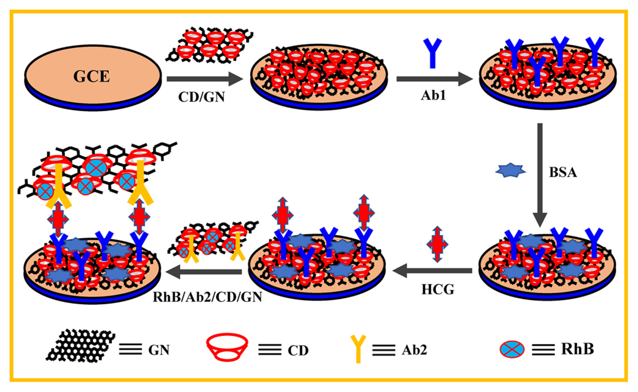
Illustrations for the construction of CD/GN-based sandwich-type electrochemical immunosensor of HCG.
2. Experimental
2.1 Reagents and Apparatus
HCG, Ab1, and Ab2 were obtained from Shanghai Lingcao Biotechnology Co., Ltd. CD, hydrazine solution, and ammonia solution were purchased from Beijing Chemical Reagent factory. Bovine serum albumin (BSA) was purchased from Sinopharm Chemical Reagent Co., Ltd. The supporting electrolyte was phosphate buffer solution (PBS) that was prepared with KH2PO4 (0.1 M), K2HPO4 (0.1 M) and KCl (0.1 M). CHI 660E Electrochemical Workstation was applied for the electrochemical measurements. GCE (with a diameter of 3 mm), platinum wire and Ag/AgCl electrode were used respectively as the working electrode, counter electrode and reference electrode.
2.2 Preparation of CD/GN
Firstly, graphene oxide (GO) was prepared with the typical Hummers method [23]. For preparing CD/GN, the homogeneous GO dispersion of 50.0 mL (1.0 mg mL−1) and 50.0 mL CD dispersion (1.0 mg mL−1) were mixed in a glass flask to be stirred continually for 15 min before the addition of ammonia solution and hydrazine solution. Then, the formed mixture solution was ultrasonicated for 30 min and subsequently heated at 65°C for 8 h under stirring. Finally, the CD/GN nanohybrid was obtained after filtration and drying.
2.3 Construction of RhB/Ab2/CD/GN
For constructing RhB/Ab2/CD/GN, 1.0 mL CD/ GN dispersion (0.25 mg) was mixed with 9.0 mL Ab2 solution (200 μg mL−1) and 0.1 mL RhB solution (0.2 mg mL−1), then the mixture was vibrated at 4°C for 5 h. Finally, the obtained RhB/Ab2/CD/GN was washed and stored for further use.
2.4 Construction of sandwich-type immunosensor
Firstly, GCE was modified with 8.0 μL CD/GN suspension (0.25 mg mL−1) and dried. Next, 3.0 μL Ab1 (0.2 mg/mL) was incubated on the surface of CD/GN/GCE. For preventing the non-specific binding, Ab1/CD/GN/GCE was incubated in BSA solution. Next, the formed Ab1/CD/GN/GCE was interacted with HCG target and then HCG-Ab1/CD/ GN/GCE was immersed in the RhB/Ab2/CD/GN solution to construct a sandwich-type electrochemical immunosensing platform. The electrochemical impedance spectroscopy (EIS) analysis was carried out in 5.0 mM [Fe(CN)6]3−/4− solution, and the related experimental setup were as follows: initial potential, 0.25 V; low frequency, 1 Hz; high frequency, 1000000 Hz; amplitude potential, 0.005 V.
3. Results and Discussion
3.1 Characterization of CD/GN
Various technologies were introduced to characterize the as-prepared CD/GN nanohybrid. Firstly, Fig. 1a–d showed the atomic force microscopy (AFM) images of GN and CD/GN, the results revealed that the average thickness of GN is ~0.5 nm. As for the CD/GN nanohybrid (Fig. 1b and d), the average thickness increases to be ~1.2 nm owing to the presence of CD molecules on the surface of GN. Then, the FT-IR spectrums of GN and CD/GN were displayed in Fig. 1e, it can be found that the pure GN exhibits typical C-C band (1132 cm−1) and C=C conjugation (1565 cm−1) [24–26], while the synthesized CD/GN shows several typical CD absorption spectrums: the ring vibrations at 482, 622, and 754 cm−1, the C-O/C-C stretching/O-H vibrations at 1029 and 1085 cm−1, the CH2 stretching vibrations at 2915 cm−1, and the O-H stretching vibrations at 3423 cm−1 [26,27]. In addition, the dispersibility of CD/GN in water was also studied and the images of GN and CD/GN were shown in Fig. 1f. From the images, it can be found that the pure GN exhibits poor dispersibility, while the CD/GN exhibits superior dispersibility in water resulted from the hydrophilic exterior of CD. These characterizations indicate that the CD/ GN nanohybrid was prepared successfully and the CD molecules can improve greatly the dispersibility of GN.
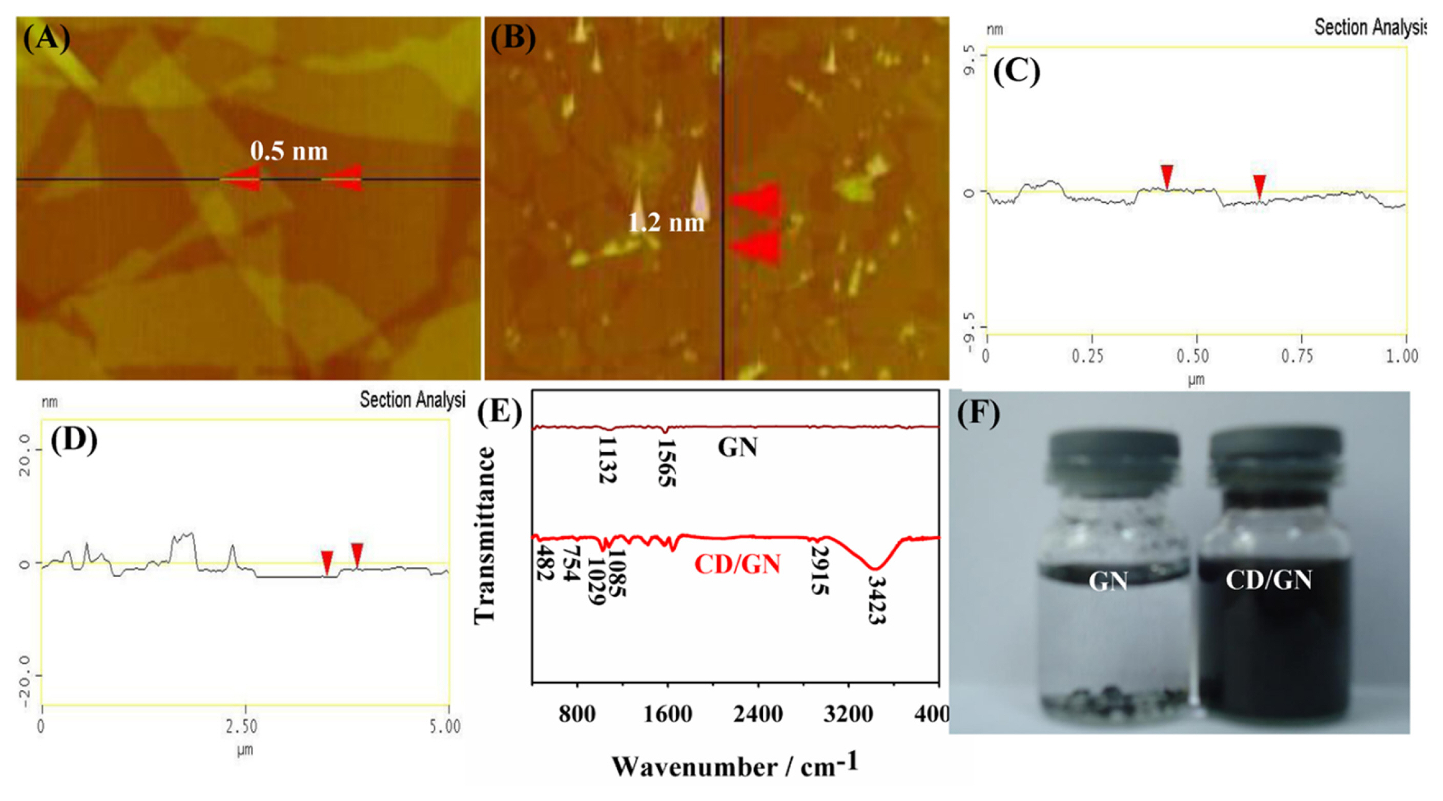
AFM images and the height profiles along the lines of (a,c) GN and (b,d) CD/GN; (e) FT-IR spectrums and (f) photographs of dispersions of GN and CD/GN.
3.2 Electrochemical immunosensing for HCG
Firstly, the EIS analysis was used to investigate the construction processes of the sandwich-type immunosensor, and the EIS plots of CD/GN/GCE, Ab1/ CD/GN/GCE, HCG-Ab1/CD/GN/GCE and RhB/ Ab2/CD/GN-HCG-Ab1/CD/GN/GCE in 0.1 M KCl solution containing 5.0 mM [Fe(CN)6]3−/4− were shown in Fig. 2. The results revealed that the charge-transfer resistance (RCT) value of CD/GN/GCE is extremely small owing to the prominent conductivity of CD/GN. However, as for the other electrodes including Ab1/CD/GN/GCE, HCG-Ab1/CD/GN/ GCE and RhB/Ab2/CD/GN-HCG-Ab1/CD/GN/ GCE, the Rct values at these electrodes increased in turn, the reason is that the conductivities of biomacromolecules including Ab1, BSA, HCG and Ab2 are not as good as that of CD/GN. This investigation revealed that the sandwich-type immunosensor layers were constructed successfully.

The EIS plots of (a) CD/GN/GCE, (b) Ab1/CD/GN/ GCE, (c) HCG-Ab1/CD/GN/GCE and (d) RhB/Ab2/CD/ GN-HCG-Ab1/CD/GN/GCE.
Next, the feasibility of the designed sandwich-type immunosensor for HCG was demonstrated by measuring the differential pulse voltammetry (DPV) responses of different electrodes. As exhibited in Fig. 3, the results showed that no oxidation peak current of RhB presented at HCG-Ab1/CD/GN/GCE in 0.1 M PBS (pH 7.0) due to the absence of RhB/Ab2/CD/ GN. However, there is an obvious peak current of RhB presented at ~0.855 V for RhB/Ab2/CD/GN-HCG-Ab1/CD/GN/GCE since the antibody-antigen recognition reaction between Ab2 and HCG (6.0 ng mL−1) induced the capture of RhB/Ab2/CD to HCG-Ab1/ CD/GN/GCE. For further comparison, the sensor performances without the use of CD have been studied. As exhibited in Fig. 3, there is only an incredibly small peak current observed that can even be ignored at RhB/Ab2/CD/GN-HCG-Ab1/GN/ GCE, indicating the non-specific adsorption is extremely weak. These comparisons revealed that the as-designed immunosensing proposal is feasible for the HCG detection.
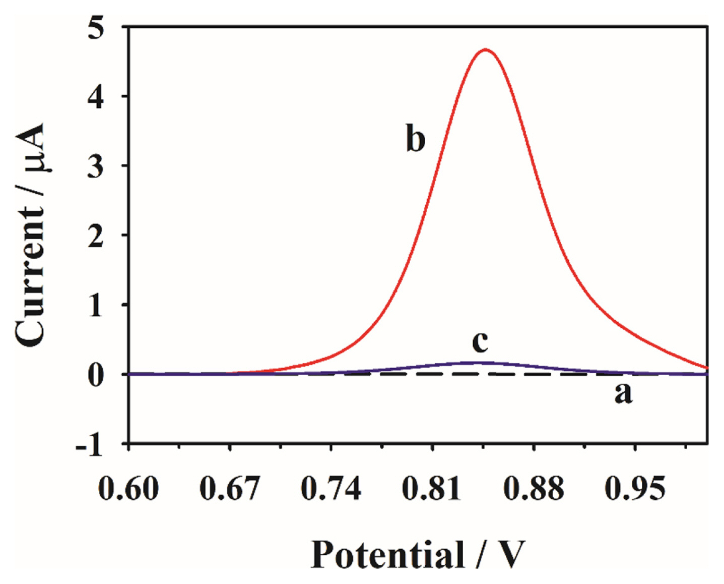
The DPV response of (a) HCG-Ab1/CD/GN/GCE, (b) RhB/Ab2/CD/GN-HCG-Ab1/CD/GN/GCE and (c) RhB/Ab2/CD/GN-HCG-Ab1/GN/GCE in 0.1 M PBS.
For achieving the highest sensitivity in detecting HCG, several related experimental conditions were evaluated then (HCG, 6.0 ng mL−1). Fig. 4A exhibited the influence from the different pH values of PBS on the peak current of RhB/Ab2/CD/GN-HCG-Ab1/ CD/GN/GCE, it’s noted the DPV response of the immunosensor increases along with the pH increase from 5.0 and reaches a maximum at 7.0, the reason may be that the excessive acid or alkaline conditions destroy the protein structures. Fig. 4B showed the influences from the interaction time of Ab1/CD/GN with HCG and HCG with RhB/Ab2/CD/GN (HCG, 6.0 ng mL−1; pH 7.0), the results revealed that the optimum time for the former is 32 min, while 40 min is for the latter, thus these times were used for the antigen--antibody interactions. In addition, the amount of Ab2 used in constructing RhB/Ab2/CD/ GN was also investigated and the results were shown in Fig. 4C. From the figure, it can be seen that the DPV current of the immunosensor increases gradually with the increase of the amount ranging from 3.0 to 9.0 μL. However, when the amount of Ab2 increases further, the DPV current showed a decrement owing to the competitive immobilization between Ab2 and RhB probe with CD, revealing the ideal balanced point between RhB and Ab2 is 9.0 μL.

The effects of (A) pH values of PBS, (B) interaction time of (a) Ab1/CD/GN with HCG and (b) HCG with RhB/Ab2/CD/GN, and (C) the used amount of Ab2 for RhB/Ab2/CD/GN.
Finally, under the conditions optimized above, the detection capability of the designed electrochemical immunosensor for HCG was evaluated. As displayed in Fig. 5, via plotting the calibration curve that consisted of the DPV currents of RhB upon the concentrations of HCG, the obtained results revealed that the currents increase linearly along with the increase of HCG concentrations ranging from 0.002 and 10.0 ng mL−1, and the regression equation is Ip(mA) = 0.304 + 0.686C (ng mL−1) (R2 = 0.997). Herein, C is expressed the concentration of HCG. Based on S/N = 3, the calculated limit of detection (LOD) is low to 1.0 pg mL−1. For understanding the detection capability of the immunosensor more direct-viewing, the comparisons between the present work and the previous literatures for the analytical parameters including linear range and LOD were performed, and the results of comparison were shown in Table 1. From the comparisons, it can be known that the detection performances of the designed electrochemical immunosensor are more superior (wider linearity and lower LOD) than those at the most of previous literatures.
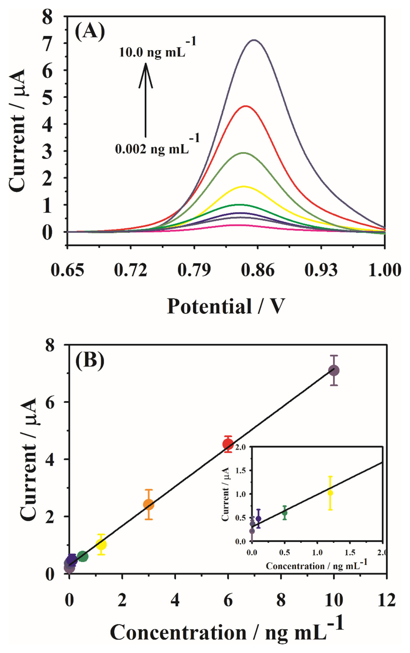
(a) DPV responses of the immunosensor at different concentrations of HCG from 0.002 ng mL−1 to 10.0 ng mL−1; (b) the related calibration plot for HCG detection, the inset is the calibration curve at low concentrations.
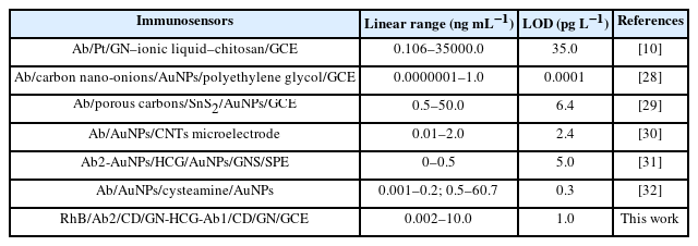
Comparisons of the designed immunosensor with the other electrochemical sensors for HCG
3.3 SSRR analysis
The SSRR performances (selectivity, stability and reproducibility as well as real application ability) are also very important for the sensors and they were studied in the following sections with the concentration of 2.5 ng mL−1 for HCG. Firstly, the selectivity of the designed immunosensor was performed that was carried out in the presence of several interfering substances (Fig. 6a). The results revealed the peak currents of RhB did not change even in the presence of cortisol (CT), alpha fetoprotein (AFE), dehydroepiandrosterone (DPD), carcinoembryonic antigen (CEA), leptin (LT), BSA, and haptoglobin (HG) (10.0 times of HCG level, that is 25.0 ng mL−1), demonstrating that the designed sandwich-type immunosensor exhibited desirable selectivity for HCG detection. Next, the stability of the immunosensor was studied, and the test results showed that the signal responses of the immunosensor remain almost unchanged for four weeks through storing RhB/Ab2/ CD/GN at 4? before use (Fig. 6b), suggesting the developed immunosensor offered satisfactory stability. Then, we also investigated the reproducibility of the proposed immunosensor by recording the peak current responses of RhB at 8 constructed independently RhB/Ab2/CD/GN-HCG-Ab1/CD/GN/ GCE. As shown in Fig. 6c, the obtained relative standard deviation (RSD) value at these immunosensors is 3.65%, indicating the reproducibility of the present proposal was acceptable. Furthermore, the real application capability of the proposal was also studied by detecting HCG in real human serum via standard addition method. The serum sample was obtained from the local hospital and various concentrations of HCG were added in turn to the samples, sequentially the signal currents of the sensing platform towards the sample were recorded, and the recovery was used to evaluate the feasibility of immunosensor in real application. As displayed in Table 2, the obtained recoveries were noted in the range between 95.0 and 98.0%, revealing the designed sandwich-type immunosensor has desirable real application capability for HCG analysis.
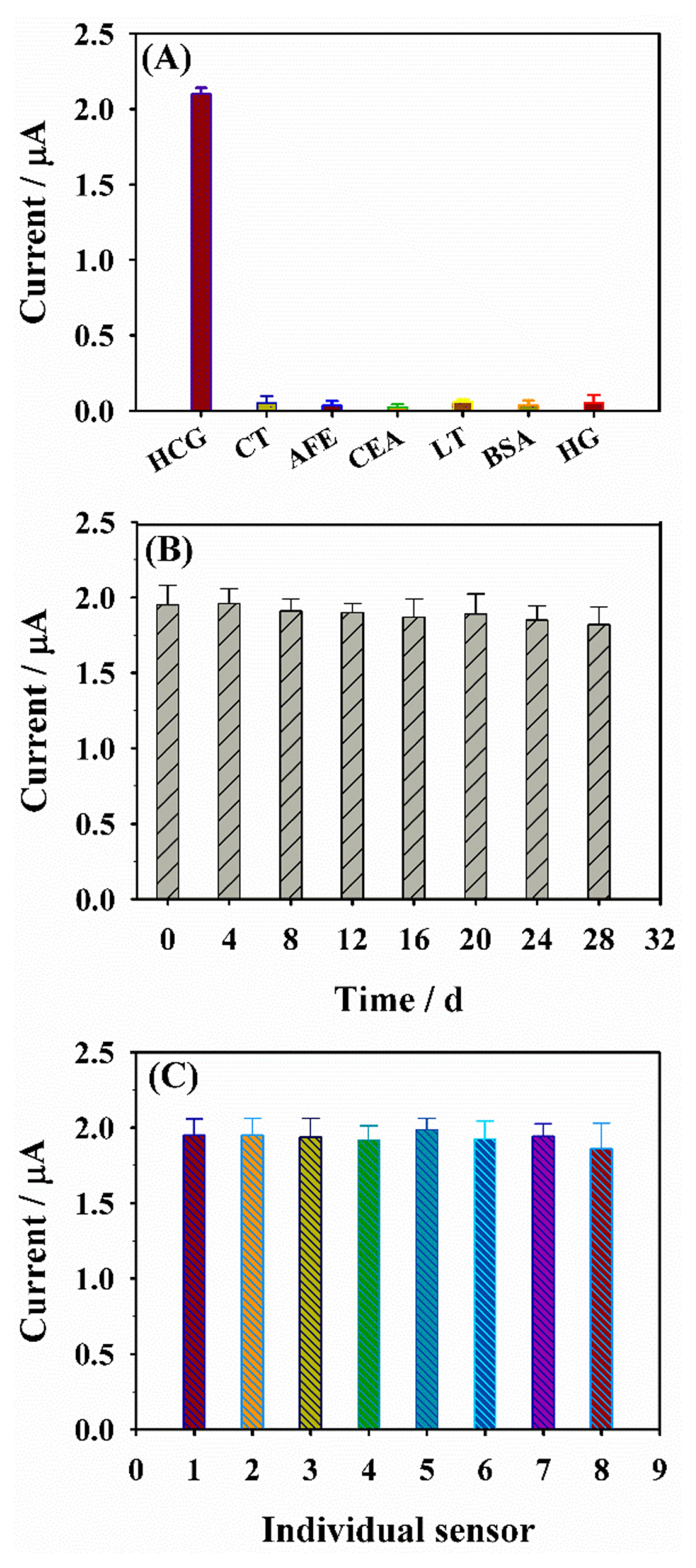
(a) Selectivity, (b) stability and (c) reproducibility for HCG detection based on the sandwich-like immunosensor.

The detection of HCG in human serum sample
4. Conclusions
In summary, a simple and sensitive electrochemical sandwich-type immunosensor was designed for HCG in this work by synthesizing CD/GN nanohybrid as simultaneously sensing platform and signal transducer coupled with RhB as signal probe. After optimizing several key parameters, the designed sandwich-type immunosensor can offer superior analytical performances including wide linear range of 0.002–10.0 ng mL−1 and low LOD of 1.0 pg mL−1, which are more superior than most of previous works. Furthermore, the designed immunosensor exhibited desirable selectivity, stability and reproducibility as well as real application ability, it’s hence confirmed the designed simple and sensitive proposal for the HCG determination has great potential application.
Acknowledgment
The work was financially supported by Fuzhou health technology project (2019-S-wp7).